What is Macular Hole?
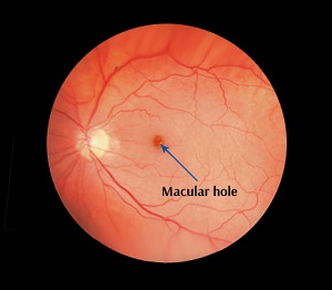
A macular hole is a defect in the macula, the central part of the retina (light receptive tissue of the eye). The macula is responsible for the sharp central vision required for reading, driving, etc. The central part of the macula called the fovea provides the best possible sharpest vision and is the part of the eye affected by macular holes.
In the early stages, a macular hole causes distorted or blurred vision and straight lines may appear bent or wavy. You may have difficulty with reading and other work that requires seeing detail. As the condition progresses, you can see a blind spot (small dark spot) in the center of the visual field.
Macular holes are more common in women than men and usually occur in people over 50 years old due to anatomical changes that occur in the eyes with aging. The vitreous, a gel-like substance that fills the center of the eye, is firmly attached to the retina, but with aging, the vitreous gel shrinks and pulls away from the retina causing a macular hole.
Other factors that can cause a macular hole include severe near-sightedness, blunt injury to the eye, severe swelling of the retina or its detachment from the underlying layers of tissue.
Three stages to a macular hole based on its size and progression
- Stage 1 (foveal detachments) - If left untreated, about 50% of stage 1 macular holes will progress.
- Stage 2 (partial-thickness holes) - If left untreated, 70 percent of holes will progress.
- Stage 3 (full-thickness holes) - affect central and detailed vision. If left untreated, a stage 3 macular hole can lead to detachment of the retina from the underlying layers of tissue.
Diagnosis of Macular Hole
To diagnose a macular hole, the doctor will instill eye drops and dilate your pupils to view the retina. A test called fluorescein angiography is performed that uses a special dye to illuminate areas of the retina. Another accurate diagnostic test is the optical coherence tomography (OCT) which uses a laser camera to capture pictures of the retina in which small macular holes that cannot be identified in a Fluorescein angiography can be detected.
If the macular hole is very small and does not cause a major impact on vision, treatment may not be required, but regular eye examinations are recommended to ensure the macular hole does not enlarge or have other effects on the eye. However, in many cases, surgery is required to improve vision. Vitrectomy is the most effective surgery for a macular hole.
Related Topics
- Retinal Detachment
- Retinal Tear
- Diabetic Retinopathy
- Age-Related Macular Degeneration
- Retinal Vascular Diseases
- Retinal Artery Occlusion
- Retinal Vein Occlusion
- Retinal Hemorrhage
- Vitreous Hemorrhage of any Etiology
- Central Serous Retinopathy
- Posterior Vitreous Detachment
- Vitreomacular Traction Syndrome
- Epiretinal Membrane
- Macular Edema
- Macular Hole
- Retinoschisis
- Ocular Ischemic Syndrome
- Cystoid Macular edema
- Color Blindness
- Nyctalopia/Night Blindness
- Cone Dystrophy
- Choroideremia
- Retinopathy of Prematurity
- Uveitis & Ocular Inflammation
- Retinoblastoma